

What Nutrients People are deficient in a Gluten-Free Diet? | Supplements that Help to Improve Gluten Intolerance Conditions
Is Gluten Free Diet for you?
The spectrum of gluten-related disorders includes celiac disease, dermatitis, gluten ataxia (Severe body's reaction to gluten impacting central nervous system), wheat allergy and non-celiac gluten sensitivity. The term non-celiac gluten sensitivity (NCGS) describes a clinical condition when the individuals develop symptoms to gluten-containing foods and feel better on the absence of gluten but do NOT have celiac disease.

What is gluten?
Gluten is the Latin word for glue. Gluten is not a single protein but instead a complex combination of proteins, primarily gliadin and glutenin. Gluten is formed in the process of dough making when wheat flour is mixed with water. The combination of gliadin, a viscous (thick) protein, with glutenin, a long, elastic protein, yields gluten with its unique “visco-elastic” properties that are important in baking and food processing. Therefore despite the common interpretation of the term “gluten-containing grains,” in fact, gluten itself is not present in the wheat seed; rather it is created during the process of dough making.
Furthermore, Rye and barley, do not form gluten. These grains contain proteins known as secalin in rye and hordein in barley which both contain fragments acting similar to wheat's gliadin in the body triggering celiac disease.
All three grains—wheat, rye, and barley—are commonly and by error considered “gluten-containing grains”, just for simplicity and convenient.
Protein compounds of Gliadin (wheat), secalin (rye), and hordein (barley) are rich in two amino acids including proline and glutamine, and thus are called together “prolamins”.
The chemical bonds between proline and glutamine in the gluten protein are resistant to digestion. This prevents complete breakdown of gluten into small, harmless molecules. Instead, big, undigested gluten fragments remain in the digestive tract.
In healthy people, these large, undigested fragments of gluten protein are eventually harmlessly excreted and do not provoke an immune response (Fasano 2009). For people with celiac disease, however, these glutamine-proline rich fragments, or peptides, are toxic.

What Are The Differences Between Wheat Allergy, Celiac Disease, and Non-Celiac Gluten Sensitivity?
While clinical symptoms of these conditions may overlap and make it difficult to differentiate them on the basis of symptoms alone, these disorders are actually three different conditions from distinct processes:
In wheat allergy certain type of antibody (IGE) is produced in reaction to wheat, resulting in the release of histamine and other inflammatory compounds which could cause symptoms such as skin eruptions, runny nose, itching, and in rare cases, anaphylactic reaction.
Wheat allergy is a less common condition in about 0.5% of the general population. IgE antibodies are key players in immediate food allergy reactions which usually occur within minutes to several hours after ingesting the allergenic food, though the reaction can occur up to two days later IgE reactions to foods can be measured by blood or dermal testing. (Available by Health Palace)
Celiac disease is an autoimmune disease, not an allergy. Autoimmune disease reactions are long lasting and highly destructive, whereas allergic reactions can appear and disappear within minutes to hours after contact with an allergen. In celiac disease other than signs and symptoms, there is a strong presence of antibody IGA in the blood sample with positive villous atrophy (damage to small intestine) on biopsy, and positive human leukocyte antigens (HLAs). Blood tests alone are often not considered adequate to confirm a diagnosis of celiac disease. More than 98% of people with celiac disease have at least one of two variants of a gene called HLA DQ2 and/or HLA DQ8 .
Genetic testing for HLA DQ2 and HLA DQ8 is useful due to several reasons; negative tests rule out almost all cases of celiac disease, including those already on a gluten-free diet; negative tests may help clarify the picture when the result of other testing is unclear; and these tests are used to screen close relatives of patients for genetic risk of celiac disease . (available at Health Palace).
Celiac disease is an inflammatory immune disorder in genetically susceptible individuals. In people with celiac disease, ingestion of gluten provokes an immune attack that inflames and damages the lining of the small intestine. Gluten causes damage to the gut of individuals who suffer from celiac disease. This typically results in nutrient malabsorption along with a wide variety of symptoms, ranging from diarrhea and constipation to skin rashes and depression . This reaction to gluten is not an allergy, but rather an internal inflammatory immune condition. Untreated celiac disease contributes to variety of other conditions such as osteoporosis, infertility, neurological disorders, other autoimmune diseases, and in some cases, cancer.
Celiac disease is characterized by increased intestinal permeability (“leaky gut”), which allows gluten to get through the damaged gut barrier and provoke an immune response.
In celiac disease, intestinal damage manifests as a flattening of the villi (finger shape structures of the intestinal cells which are significantly important for the absorption of nutrients ). The villous atrophy reduces the surface area available for nutrient absorption resulting mal-absorption which further contributes to both intestinal disorders such as indigestion and diarrhea, and non-intestinal conditions such as anemia and osteoporosis.
Celiac disease has been associated with a number of other conditions, especially autoimmune-related conditions such as type 1 diabetes and autoimmune thyroid disease, and an increased risk of certain cancers such as non-Hodgkin lymphoma, small intestinal cancer, colon cancer, and basal cell carcinoma.
Brain and nervous system problems are seen in celiac cases. In celiac disease autoimmune antibodies are produced against and enzyme called transglutaminase involving the intestine and skin, however a sub type of this enzyme called transglutaminase 6, is found in the brain and nervous system. Antibodies against this enzyme have been identified in patients with gluten ataxia and schizophrenia. Although, the involvement of these antibodies in other neurological and psychiatric conditions is still under investigation, there is evidence that transglutaminase 6, and the transglutaminase enzyme family in general, may be important in the development of degenerative diseases of the brain. For example, Recently, the prevalence of celiac disease was reported to be 5–10 times higher in a study in patients with multiple sclerosis compared to the general population. All the patients in this study had an excellent response to the gluten-free diet in an average of three years, with regard to both digestive and neurological symptoms.
A case report has shown celiac disease can mimic multiple sclerosis. A patient with a history of diarrhea and colic since youth and neurological symptoms since age 18 was diagnosed with multiple sclerosis at age 34. Later was found the he was positive for HLA DQ2 and DQ8 genetic tests, and under a gluten-free diet both his digestive and neurological symptoms have been improved.

Non-celiac gluten sensitivity (NCGS) is only considered once wheat allergy and celiac disease have been ruled out, and a beneficial response to dietary elimination of gluten-containing foods is observed. Specific antibodies are absent and there is no villous atrophy.
Majority of the cases of Non-celiac gluten sensitivity, the symptoms are reported to the food that contained gluten, not the pure gluten; therefore, suggesting this group are may be actually sensitive to other compounds known as FODMAPs rather than gluten.
FODMAPs (fermentable oligosaccharides, disaccharides, monosaccharides, and polyols) are types of carbohydrates that some people cannot digest very well. So the bacteria in the colon ferment these carbohydrates resulting in gas, bloating, abdominal pain and diarrhea. Wheat, barley and rye are high in FODMAP, which may be a contributing factor to these symptoms. Not all carbohydrates are considered FODMAPs, but many types of foods contain them, including foods that are high in fructose, like honey, apples, mangoes, and watermelon; dairy products, like milk and ice cream; and fructans, such as garlic and onions.
In fact, FODMAPs seem more likely than gluten to cause widespread intestinal distress, since bacteria regularly ferment carbohydrates but ferment protein less frequently. Although a FODMAP-free diet is complicated, it permits people to eliminate individual foods temporarily and then reintroduce them systematically to determine which, if any, are responsible for their stomach problems. Furthermore, wheat contains other proteins called amylase/trypsin inhibitors (ATI) that in lab has shown to cause intestinal inflammation.
People with NCGS can experience a spectrum of highly variable symptoms similar to those caused by celiac disease and irritable bowel syndrome such as bloating, abdominal pain, diarrhea, constipation, nausea, acid reflux, and mouth ulcers . These Individuals may have non-intestinal symptoms such as feeling generally unwell, fatigue, headaches, foggy mind, numbness, joint pains, or skin rash. An individual may have one or more symptoms.
Non-celiac gluten sensitivity does not appear to be an autoimmune condition, making it distinct from celiac disease; neither is an allergic condition, which distinguishes it from classical wheat allergy. In fact, there are currently no universally recognized tests or biomarkers for non-celiac gluten sensitivity. A recent randomized controlled trial in 37 patients with NCGS and irritable bowel syndrome found that gluten did not produce negative effects when FODMAPs were restricted in the diet.
What Is A Gluten Free Diet?
A strict, lifelong gluten-free diet is the only established treatment for celiac disease, as even a tiny amount of gluten can lead to intestinal damage in those with celiac disease. In the majority of cases, a strict compliance has helped to reduce their symptoms, to heal the existing intestinal damage, and to prevent further damage.
The gluten-free diet excludes all foods that contain wheat (including spelt, triticale, and kamut), rye, and barley. This means avoiding these grains, as well as pasta, cereals, and processed foods—unless they are labeled “gluten-free.”
It is thought that most celiac patients can eat modest amounts of pure gluten free oats, while some celiac patients do not tolerate oats well.
The gluten-free diet is also recommended for non-celiac gluten sensitivity conditions as well; however, recent studies suggests avoidance of FODMAPs as opposed to gluten. The level of gluten tolerance varies among individuals with NCGS and with rare exceptions, most of these individuals can eat trace amounts of gluten without negative health consequences.

What Can Help To Improve The Gluten Related Conditions?
Certain specific types of protease enzymes derived from plants and microorganisms such as common fungus Aspergillus niger can completely break down gluten peptides into harmless fragments. These enzymes are proline-specific and /or glutamine-specific protease. These enzyme specifically cleaves the proline-rich gliadin peptides, making them less immunogenic.
Experiments with cell cultures suggests that these enzymes are resistant to the strong acidic environment of the stomach, and fully digest gluten and immunogenic peptides and appeared to reduce gluten-induced small intestinal damage in patients with celiac disease who were challenged with up to 2g gluten per day while following a gluten-free diet.
Low pancreatic digestive enzymes, is relatively common in celiac disease patients. Damage to the small intestine impacts on the ability of the pancreas to secrete digestive enzymes. Patients treated with pancreatic enzymes experienced a 75% reduction in symptoms. Another study found that the severity of villous atrophy in the small intestine was greatly associated with laboratory signs of pancreatic enzyme insufficiency.
Celiac patients and those on a gluten free diet due to poor intestinal absorption of nutrients, chronic intestinal inflammation, limited diet, and limited choices due to cross contamination with gluten byproducts, are prone to develop many health conditions related to long term deficiencies of essential nutrients. These conditions often increase the severity of the digestive conditions, despite of being on a restrict diet.
Poor calcium absorption is very common in individuals with celiac disease due to damage to the intestinal lining . This chronic deficiency leads to bone loss, osteopenia, osteoporosis, and increased fracture risk. A calcium deficient gluten free diet is a major cause of low bone mineral density. Therefore, supplementation with calcium is very important for patients with celiac disease or those on the gluten free diet.
Several studies indicate that the celiac Patients and those on the gluten free diet are more at risk of magnesium deficiency as well. Magnesium is an essential mineral involved in many of enzymatic reactions in the body and it is required for proper metabolism of vitamin D and calcium, and therefore for bone health. The significance of the magnesium deficiency in celiac disease was first recognized over fifty years ago. And in later studies it was found that children and adolescents with celiac disease or normal intestinal villi who had been on a gluten-free diet were depleted form magnesium. Taking Magnesium supplements in these individuals resulted in increases in both red blood cell magnesium and bone mineral density. Accordingly, screening for magnesium status and deficiency, and magnesium supplementation and dietary enrichment, are considered essential aspects of care for celiac patients and those on a gluten free diet.
Based on studies 64% of men and 71% of women with celiac disease and on a gluten free diet are found to have significantly low vitamin D levels contributing to range of musculoskeletal disorders including bone pain, muscle disorders (myopathy), loss of bone mineral density, osteopenia, and osteoporosis. Vitamin D supplementation helps to improve the symptoms and prevent deficiencies. Vitamin D also plays important role to particularly modulate the immune system and provides anti-inflammatory benefits. Supplementation with vitamin D and calcium is widely recommended for celiac patients. Lab studies suggests vitamin D potentially decrease systemic inflammation and prevent autoimmune disease in human.
B-deficiency and anemia related to low b12 and folate is very common in celiac cases and on a gluten free diet. Low levels of B12, folate and B6 are also associated with elevated homocycteine levels, which contributes to a damage to lining of blood vessels and promote atherosclerotic disease. Vitamins B12, B6, and folate, are necessary for the metabolism of homocysteine, and their supplementation is shown to help normalize the homocycteine levels and prevent deficiencies in celiac conditions and in individuals on a gluten free diet.
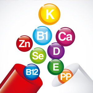
Celiac individuals and those on a poor gluten free diet are prone to deficiency of fat soluble vitamins and nutrients including vitamin A, D, E, K, and omega 3s.
Poor nutrient absorption in celiac condition is associated with vitamin A deficiency. Vitamin A is important for normal immune function, vision, and gene expression. Case studies have pointed out the importance of vitamin A, even in remitted celiac cases on a gluten-free diet. Supplementing with Vitamin A has helped to restore their intestinal symptoms and blurry vision.
Vitamin E is an important fat-soluble vitamin and free radical scavenger that plays a major role in protecting cell membranes from oxidative damage. Vitamin E deficiency has also been linked to the development of neurological symptoms in celiac patients.
Because often patients on the gluten free diet do not get adequate amount of calcium, vitamins K and D, supplementation with vitamin K at the time of diagnosis for these individuals has been recommended. Vitamin K participate in calcium metabolism and therefore it is important for bone health. Furthermore, Vitamin K deficiency can contribute to easy bruising, which is a atypical sign of celiac disease.
Serum concentrations of omega-3 fatty acids are significantly lower in celiac individuals and after one year on a gluten-free diet, levels of omega-3 fatty acids still remaine well below control values. Omega-3 fatty acids significantly contribute to biological anti-inflammatory effects in the body.
In vitro studies on the intestinal epithelial cells cultures suggests that supplementation with DHA would help to reduce intestinal inflammation. Intestinal cells in response to inflammation triggered by gluten, release inflammatory arachidonic acid, while DHA is shown to be able to block its release. Also, omega-3 fatty acids help reduce inflammation by inhibiting the nuclear factor-kappaB (NF-ĸB) which is a pro-inflammatory mediator. Omega 3 is able to stimulate PPAR- Gamma cellular receptor which activates genes that reduce the production of pro-inflammatory markers.
Celiac individuals and those on a gluten free diet are at the higher risk of developing autoimmune thyroid conditions, due to selenium deficiency. Selenium is an integral part of the enzyme glutathione peroxidase which the body's master antioxidant; plus, Selenium is essential for thyroid hormone regulation and conversion.
Glutathione is our body's powerful defenses against free radicals, and is essential to the liver’s detoxification functions. Celiac patients are prone to lower levels of glutathione in their intestinal lining and blood, resulting in elevated lipid peroxides (oxidized fat) which promotes and contributes to oxidative damage to the intestinal tissue and increased risk of cancer. Supplementation with glutathione has shown to help to restore the body's reserve. Also, N-acetyl cysteine as well as alpha-lipoic acid, vitamin c, selenium, and whey protein are helping to improve and maintain proper glutathione level and functions.
Alpha-lipoic acid is naturally produced in the mitochondria of the cell contributing to energy production. It is also a powerful free radical scavenger, and it helps reviving other antioxidants such as glutathione, vitamin C, vitamin E, and coenzyme Q10. Dietary supplementation with alpha-lipoic acid is shown to increase both cellular glutathione concentrations and its activity.
Zinc deficiency is another common condition associated with celiac disease and gluten free diet. Zinc is an essential nutrient which participates in more than 300 enzymatic reactions in the body. Some consider the zinc supplementation a necessary part of celiac treatment and an addition to the gluten free diet. Zinc deficiency negatively impacts growth, immune function, wound healing, and skin problems.
Zinc carnosine in particular appears to help maintain the integrity of the intestinal barrier and inhibit the intestinal permeability in the treatment group.
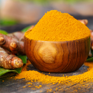
Since celiac is an inflammatory autoimmune condition, keeping the inflammation and the inflammatory markers at the low level, helps to improve symptoms and the quality of life of those individuals. Anti-inflammatory nutraceuticals such as Curcumin and Boswellia serrata are shown to help with variety of inflammatory conditions including gluten intolerance or celiac disease. Curcumin contributes to suppression of interferon-gamma, an inflammatory marker; while boswellia participates in inhibition of pro-inflammatory pathways involving 5-lipoxygenase, leukotrienes, and TNF-α which both In a similar way, may help reduce the intestinal inflammation in celiac disease.
EGCG a chief constituent of Green tea extract is capable of interrupting inflammatory tissue damage seen in celiac and other autoimmune conditions. Helper T cells are considered a key factor in many autoimmune diseases including celiac disease. EGCG helps to suppress the auto-reactive T cells which consequently reduces production of pro-inflammatory cytokines. Furthermore, EGCG is shown to inhibit interferon-gamma which is a main inflammatory mediator in celiac disease .
Glutamine is the most abundant amino acid in the body and play a key role in protection and repair of the gastrointestinal lining, especially during stress and illness. Oral supplementation with glutamine helps to improve the size of intestinal villi, encourages proliferation of the cells of the intestinal lining, and helps to maintain integrity of the gut lining, prevent excessive permeability, and improve gut barrier function.
Whey protein is source of essential amino acids and it is rich in cysteine which is used to produce glutathione. Whey protein is highly effective for increasing glutathione levels in different types of cells. Whey protein also helps to significantly reduce intestinal permeability. This benefit of whey on gut integrity may be useful in celiac disease, and could be considered to be used in conjunction with gluten-free diet as a source of high-quality protein and glutathione precursors.
Evidently the intestinal micro-biome is greatly altered in celiac individuals and those on a gluten free diet. Probiotics from Lactobacillus and Bifidobacterium Species have shown to improve indigestion, constipation, and acid reflux. Probiotics help to enforce the intestinal barrier, as well as helping to predigest the gluten protein in food. Exposure of the intestinal biopsy of celiac patients to predigested gluten showed to result in less recruitment of immune cells suggesting that the probiotic formulation helped to reduce or eliminate the toxicity of wheat gluten.
Dietary & Lifestyle Changes:
What is most important for the individuals and health care professionals to know is that the diagnosis of non-celiac gluten sensitivity should not be made without excluding celiac disease. A gluten-free diet should NOT be initiated without a proper clinical assessment such as testing for IgA-tissue transglutaminase antibody while the person is on a regular gluten-containing diet, along with a confirming genetic test.
The gluten free diet can be very challenging to follow. In addition, there are concerns about the nutritional adequacy of general gluten free products on the market as they can be high in fat and sugar, and often low in fiber, iron and B vitamins. Thus those requiring or like to initiate a gluten free diet should consult with health care professionals and consider taking nutritional supplements to prevent deficiencies and to help improve their condition.
There are many gluten-free alternatives to wheat such as; amaranth, buckwheat, corn, millet, rice, quinoa, sorghum, teff, yucca, potato, nuts, flax, and all beans and legumes are naturally gluten-free. There are also alternatives to gluten-containing flour, including potato, rice, soy, tapioca, carob, and bean flour.
Gluten free foods usually contain Ingredients like rice starch, cornstarch, tapioca starch, and potato starch which are often used as replacements for white flour. But they are highly refined carbohydrates, and release so much sugar into the bloodstream.
While the gluten-free diet is also used to treat non-celiac gluten sensitivity, the need for strict adherence or permanency is not clear.
Enzyme therapy is one of the a promising intervention to detoxify gluten. Certain protein-digesting enzymes (proteases) derived from plants and microorganisms can completely break down gluten peptides into harmless fragments.
Articles and products featured by Health Palace are collected from a variety of sources and are provided as a service by Health Palace. These newsletters, while of potential interest to readers, do not necessarily represent the opinions nor constitute the advice of Health Palace. Presented materials are only for information purposes and do not intent to treat, cure, or prevent any disease.
Related Articles:
How Digestive Enzyme Can Improve Your Health?
Probiotics and their Several Health Benefits for Human
How to Reduce Inflammation Effectively With Natural Medicine
What is Homocysteine & How Is It Related to Cardiovascular Disease
References:
1.Volta U, Tovoli F, Cicola R, Parisi C, Fabbri A, Piscaglia M, . . . Caio G. Serological tests in gluten sensitivity (nonceliac gluten intolerance). Journal of clinical gastroenterology. Sep 2012;46(8):680-685.
2.Volta U, De Giorgio R. New understanding of gluten sensitivity. Nat Rev Gastroenterol Hepatol. 2012;9:295-299.
3.Volta U, Caio G, Tovoli F, et al. Non-celiac gluten sensitivity: questions still to be answered despite increasing awareness. Cell Mol Immunol. 2013;10:383-392.
4.Veraverbeke WS, Delcour JA. Wheat protein composition and properties of wheat glutenin in relation to breadmaking functionality. Critical reviews in food science and nutrition. 2002;42(3):179-208.
5.Mooney, P; Aziz, I; Sanders, D (2013). "Non-celiac gluten sensitivity: clinical relevance and recommendations for future research". Neurogastroenterology & Motility. 25 (11): 864–871. doi:10.1111/nmo.12216. PMID23937528.
6.Nijeboer, P; Bontkes, H; Mulder, C; Bouma, G (2013). "Non-celiac gluten sensitivity. Is it in the gluten or the grain?". Journal of Gastrointestinal and Liver Disorders. 22 (4): 435–40. PMID24369326.
7.Catassi C, Bai JC, Bonaz B, Bouma G, Calabrò A, Carroccio A, Castillejo G, Ciacci C, Cristofori F, Dolinsek J, Francavilla R, Elli L, Green P, Holtmeier W, Koehler P, Koletzko S, Meinhold C, Sanders D, Schumann M, Schuppan D, Ullrich R, Vécsei A, Volta U, Zevallos V, Sapone A, Fasano A (Sep 2013). "Non-Celiac Gluten sensitivity: the new frontier of gluten related disorders". Nutrients. 5 (10): 3839–53.
8.Vahedi K, Mascart F, Mary JY, et al. Reliability of antitransglutaminase antibodies as predictors of gluten-free diet compliance in adult celiac disease. Am J Gastroenterol. 2003;98(5):1079-1087.
9.Snyder CL, O Young D, Green PHR, et al. Celiac Disease. In: GeneReviews. Pagon RA, Adam MP, Amemiya A, et al. eds. Seattle WA: University of Washington, Seattle; 2015. <http://www.ncbi.nlm.nih.gov/books/NBK1727/>.
10.Smith DF, Gerdes LU. Meta-analysis on anxiety and depression in adult celiac disease. Acta Psychiatr Scand. 2012;Mar;125(3):189-93.
11.Rubio-Tapia A, Hill ID, Kelly CP, et al. ACG clinical guidelines: diagnosis and management of celiac disease. Am J Gastroenterol. 2013;108:656-676.
12.Marchioni Beery RM, Birk JW. Wheat-related disorders reviewed: making a grain of sense. Expert Rev Gastroenterol Hepatol. 2015;3:1-14. [Epub ahead of print]
13.Mansueto P, Seidita A, D'Alcamo A, Carroccio A. Non-celiac gluten sensitivity: literature review. Journal of the American College of Nutrition. 2014;33(1):39-54.
14.UCMC. University of Chicago Medical Center. The University of Chicago Celiac Disease Center. Celiac disease facts and figures. Available at: http://www.cureceliacdisease.org/wp-content/uploa... Updated 2014. Accessed 10/14/2014.
15.UCLA Health. Celiac Disease Program. For Patients page. Celiac vs Gluten-Sensitivity vs Wheat Allergies. Available at: http://gastro.ucla.edu/site.cfm?id=281. Accessed 3/6/2015.
16.Thomas H, Beck K, Adamczyk M, Aeschlimann P, Langley M, Oita RC, . . . Aeschlimann D. Transglutaminase 6: a protein associated with central nervous system development and motor function. Amino acids. Jan 2013;44(1):161-177.
17.Dieterich W, Ehnis T, Bauer M, et al. Identification of tissue transglutaminase as the autoantigen of celiac disease. Nat Med. 1997;3(7):797-801.
18.Tilley KA, Benjamin RE, Bagorogoza KE, et al. Tyrosine cross-links: molecular basis of gluten structure and function. J Agric Food Chem. 2001;49:2627-2632.
19.Stenman SM, Lindfors K, Venalainen JI, Hautala A, Mannisto PT, Garcia-Horsman JA, . . . Kaukinen K. Degradation of celiac disease-inducing rye secalin by germinating cereal enzymes: diminishing toxic effects in intestinal epithelial cells. Clinical and experimental immunology. Aug 2010;161(2):242-249.
20.Sapone A, Lammers KM, Casolaro V, et al. Divergence of gut permeability and mucosal immune gene expression in two gluten-associated conditions: celiac disease and gluten sensitivity. BMC Med. 2011;9:23.
21.Sapone A, Bai JC, Ciacci C, et al. Spectrum of gluten-related disorders: consensus on new nomenclature and classification. BMC Med. 2012;Feb 7;10:13.
22.Carroccio A, Mansueto P, Iacono G, et al. Non-celiac wheat sensitivity diagnosed by double-blind placebo-controlled challenge: exploring a new clinical entity. Am J Gastroenterol. 2012;107(12):1898-906
23.Staudacher HM, Irving PM, Lomer MC, Whelan K. Mechanisms and efficacy of dietary FODMAP restriction in IBS. Nat Rev Gastroenterol Hepatol. Apr 2014;11(4):256-266.
24.Barrett JS, Gibson PR. Fermentable oligosaccharides, disaccharides, monosaccharides and polyols (FODMAPs) and nonallergic food intolerance: FODMAPs or food chemicals? Therapeutic advances in gastroenterology. Jul 2012;5(4):261-268.
25.Catassi C, Bai JC, Bonaz B, et al. Non-celiac gluten sensitivity: the new frontier of gluten related disorders. Nutrients. 2013;Oct;5(10):3839-3853.
26.Tikkakoski S, Savilahti E, Kolho KL. Undiagnosed celiac disease and nutritional deficiencies in adults screened in primary health care. Scand J Gastroenterology. 2007;Jan;’42(1):60-65.
27.Caruso R, Pallone F, Stasi E, et al. Appropriate nutrient supplementation in celiac disease. Ann Med. 2013;Dec;45(8):522-31.
28.Stazi AV, Trinti B. Selenium status and over-expression of interleukin-15 in celiac disease and autoimmune thyroid diseases. Ann Ist Super Sanita. 2010;46(4):389-399.
29.Stazi AV, Trinti B. Selenium deficiency in celiac disease: risk of autoimmune thyroid diseases. Minerva Med. 2008;Dec;99(6):643-53.
30.Sapone A, Lammers KM, Casolaro V, et al. Divergence of gut permeability and mucosal immune gene expression in two gluten-associated conditions: celiac disease and gluten sensitivity. BMC Med. 2011;9:23.
31.Lee SA, Kim HJ, Chang KC, et al. DHA and EPA down-regulate COX-2 expression through suppression of NF-kappaB activity in LPS-treated human umbilical vein endothelial cells. Korean J Physiol Pharmacol. 2009;Aug;13:301-307.
32.Ferretti G, Bacchetti T, Masciangelo S, Saturni L. Celiac disease, inflammation and oxidative damage: a nutrigenetic approach. Nutrients. Apr 2012;4(4):243-257.
33.Fasano A. Zonulin and its regulation of intestinal barrier function: the biological door to inflammation, autoimmunity, and cancer. Physiological Reviews. 2011;Jan1;91(1):151-175.
34.Fasano A, Shea-Donohue T. Mechanisms of disease: the role of intestinal barrier function in the pathogenesis of gastrointestinal autoimmune diseases. Nat Clin Pract Gastroenterol Hepatol. 2005;Sep;2(9):416-22.
35.FARE. Food Allergy Research & Education. About Food Allergies. Wheat Allergy. Available at: http://www.foodallergy.org/allergens/wheat-allerg... Copyright 2015a. Accessed 3/6/2015.
36.Elfström P, Montgomery SM, Kämpe O, et al. Risk of primary adrenal insufficiency in patients with celiac disease. J Clin Endocrinol Metab. 2007;92(9):3595-8.
37.Peters SL, Biesiekierski JR, Yelland GW, et al. Randomized clinical trial: gluten may cause depression in subjects with non-celiac gluten sensitivity – an exploratory clinical study. 2014;May;39(10):1104-12.
38.Toouli J, Blankin AV, Oliver MR, et al. Management of pancreatic exocrine insufficiency: Australasian Pancreatic Club recommendations. Med J Aust. 2010;193(8):461-7.
39.Siegel M, Bethune MT, Gass J, et al. Rational design of combination enzyme therapy for celiac sprue. Chem Biol. 2006;13:649-658.
40.Gass J, Bethune MT, Siegel M, et al. Combination enzyme therapy for gastric digestion of dietary gluten in patients with celiac sprue. Gastroenterology. 2007;133:472-480.
41.Hallert C, Grant C, Grehn S, et al. Evidence of poor vitamin status in celiac patients on a gluten-free diet for 10 years. Aliment Pharmacol Ther. 2002;16:1333-1339.
42.Allred LK, Ritter BW. Recognition of gliadin and glutenin fractions in four commercial gluten assays. Journal of AOAC International. Jan-Feb 2010;93(1):190-196.
43.Evans KE, Leeds JS, Morley S, Sanders DS. Pancreatic insufficiency in adult celiac disease: do patients require long-term enzyme supplementation? Digestive diseases and sciences. Oct 2010;55(10):2999-3004.
44.Saibeni S, Lecchi A, Meucci G, et al. Prevalence of hyperhomocysteinemia in adult gluten-sensitive enteropathy at diagnosis: role of B12, folate, and genetics. Clin Gastroenterol Hepatol. 2005;3:574-580.
45.Reinken L, Zieglauer H. Vitamin B-6 absorption in children with acute celiac disease and in control subjects. J Nutr. 1978;108:1562-1565.
46.Hadithi M, Mulder CJJ, Stam f, et al. Effect of B vitamin supplementation on plasma homocysteine levels in celiac disease. World J Gastroenterol. 2009;Feb 28;15(8):955-960.
47.Dahele A, Ghosh S. Vitamin B12 deficiency in untreated celiac disease. Am J Gastroenterology. 2001;96(3):745-750.
48.Dickey W, Ward M, Whittle CR, et al. Homocysteine and related B-vitamin status in coeliac disease: effects of gluten exclusion and histological recovery. Scand J Gastroenterol. 2008;43:682-688.
49.Dickey W. Low serum vitamin B12 is common in celiac disease and is not due to autoimmune gastritis. Eur J Gastroenterol Hepatol. 2002;14(4):425-427.
50.Turner J, Pellerin G, Mager D. Prevalence of metabolic bone disease in children with celiac disease is independent of symptoms at diagnosis. J Pediatr Gastroenterol Nutr. 2009;49:589-593.
51.Szymczak J, Bohdanowicz-Pawlak A, Waszczuk E, et al. Low bone mineral density in adult celiac disease. Endokrynol Pol. 2012;63(4):270-6.
52.Pantaleoni S, Luchino M, Adriani A, et al. Bone mineral density at diagnosis of celiac disease and after 1 year of gluten-free diet. Scientific World Journal. 2014;2014:173082.
53.Lucendo AJ, Garcia-Manzanares A. Bone mineral density in adult celiac disease: an updated review. Rev Esp Enferm Dig. 2013;Mar;105(3):154-62.
54.Nikpour S. Neurological manifestations, diagnosis, and treatment of celiac disease: A comprehensive review. Iranian Journal of Neurology. 2012;11(2):59-64.
55.Bianchi ML, Bardella MT. Bone in celiac disease. Osteoporos Int. 2008;19:1705-1716.
56.Blazina S, Bratanic N, Campa AS, et al. Bone mineral density and importance of strict gluten-free diet in children and adolescents with celiac disease. Bone. 2010;Sep;47(3):598-603.
57.Rujner J, Socha J, Wojtasik A, et al. Magnesium status in children and adolescents with celiac disease. Wiad Lek. 2001;54(5-6):277-285.
58.Richards JB, Valdes AM, Gardner JP, Paximadas D, Kimura M, Nessa A, . . . Aviv A. Higher serum vitamin D concentrations are associated with longer leukocyte telomere length in women. The American journal of clinical nutrition. Nov 2007;86(5):1420-1425.
59.Prietl B, Treiber G, Pieber TR, et al. Vitamin D and immune function. Nutrients. 2013;5:2502-2521.
60.Duerksen DR. Dramatic effect of vitamin D supplementation and a gluten-free diet on bone mineral density in a patient with celiac disease. J Clin Densitom. 2012;Jan-Mar;15(1):120-123.
61.Mager DR, Qiao J, Turner J. Vitamin D and K status influences bone mineral density and bone accrual in children and adolescents with celiac disease. Eur J Clin Nutr. 2012;66:488-495.
62.Lerner A, Shapira Y, Agmon-Levin N, et al. The clinical significance of 25OH-vitamin D status in celiac disease. Clinic Rev Allerg Immunol. 2012;42:322-330.
63.Kriegel MA, Manson JE, Costenbader KH. Does vitamin D affect risk of developing autoimmune disease?: a systematic review. Semin Arthritis Rheum. 2011;40:512-531.
64.Karaahmet OZ, Unlu E, Karaahmet F, Gurcay E, Cakci A. Myopathy related to vitamin D deficiency in patient with celiac disease. Muscle & nerve. Jul 2014;50(1):147-148.
65.Agmon-Levin N, Theodor E, Segal RM, et al. Vitamin D in systemic and organ-specific autoimmune diseases. Clinic Rev Allerg Immunol. 2013;45:256-266.
66.Antico A, Tampoia M, Tozzoli R, et al. Can supplementation with vitamin D reduce the risk or modify the course of autoimmune diseases? a systematic review of the literature. Autoimmunity Reviews 2012;12:127-136.
67.Zittermann A. Effects of vitamin K on calcium and bone metabolism. Current opinion in clinical nutrition and metabolic care. Nov 2001;4(6):483-487.
68.Rastogi A, Bhadada SK, Bhansali A, et al. Celiac disease: a missed cause of metabolic bone disease. Indian J Endocrinol Metab. 2012;Sep-Oct;16(5):780-785.
69.Booth SL. Roles for vitamin K beyond coagulation. Annu Rev Nutr. 2009;29:89-110.
70.Vaynshtein G, Rosenbaum H, Groisman GM, Markel A. Celiac sprue presenting as severe hemorrhagic diathesis due to vitamin K deficiency. The Israel Medical Association journal : IMAJ. Dec 2004;6(12):781-783.
71.Higdon J. Oregon State University. Linus Pauling Institute. Micronutrient Information Center. Vitamin A. Available at: http://lpi.oregonstate.edu/infocenter/vitamins/vi... Last updated 11/2007a. Accessed 3/5/2015.
72.Alwitry A. Vitamin A deficiency in coeliac disease. The British Journal of Ophthalmology. 2000;84(9):1075-1075.
73.Higdon J. Oregon State University. Linus Pauling Institute. Micronutrient Information Center. Vitamin E. Available at: http://lpi.oregonstate.edu/infocenter/vitamins/vi... Last updated 6/2008. Accessed 3/5/2015.
74.Kleopa KA, Kyriacou K, Zamba-Papanicolaou E, et al. Reversible inflammatory and vacuolar myopathy with vitamin E deficiency in celiac disease. Muscle Nerve. 2005;31:260-265.
75.Zapata-Gonzalez F, Rueda F, Petriz J, et al. Human dendritic cell activities are modulated by the omega-3 fatty acid, docosahexaenoic acid, mainly through PPARgamma: RXR heterodimers: comparison with other polyunsaturated fatty acids. J Leukoc Biol. 2008;84:1172-1182.
76.Yan Y, Jiang W, Spinetti T, Tardivel A, Castillo R, Bourquin C, . . . Zhou R. Omega-3 fatty acids prevent inflammation and metabolic disorder through inhibition of NLRP3 inflammasome activation. Immunity. Jun 27 2013;38(6):1154-1163.
77.Westphal C, Konkel A, Schunck WH. CYP-eicosanoids – a new link between omega-3 fatty acids and cardiac disease? Prostaglandins Other Lipid Mediat. 2011;96:99-108.
78.Surette ME. The science behind dietary omega-3 fatty acids. CMAJ : Canadian Medical Association journal = journal de l'Association medicale canadienne. Jan 15 2008;178(2):177-180.
79.Calder PC. Immunomodulation by omega-3 fatty acids. Prostaglandins Leukot Essent Fatty Acids. 2007;77:327-335.
80.Calder PC. Marine omega-3 fatty acids and inflammatory processes: Effects, mechanisms and clinical relevance. Biochimica et biophysica acta. Apr 2015;1851(4):469-484.
81.Solakivi T, Kaukinen K, Kunnas T, et al. Serum fatty acid profile in celiac disease patients before and after a gluten-free diet. Scand J Gastrolenterol. 2009;44:826-830.
82.Mahmood A, FitzGerald AJ, Marchbank T, et al. Zinc carnosine, a health food supplement that stabilizes small bowel integrity and stimulates gut repair processes. Gut. 2007;56:168-175.
83.Jiang X, Dong J, Wang B, Yin X, Qin L. [Effects of organic selenium supplement on glutathione peroxidase activities: a meta-analysis of randomized controlled trials]. Wei sheng yan jiu = Journal of hygiene research. Jan 2012;41(1):120-123.
84.Higdon J. Oregon State University. Linus Pauling Institute. Micronutrient Information Center. Selenium. Available at: http://lpi.oregonstate.edu/infocenter/minerals/se... Last updated 11/2007b. Accessed 3/5/2015.
85.Zavorsky GS, Kubow S, Grey V, et al. An open-label dose-response study of lymphocyte glutathione levels in healthy men and women receiving pressurized whey protein isolate supplements. Int J Food Sci Nutr. 2007;58(6):429-436.
86.Stojiljkovic V, Todorovic A, Radlovic N, et al. Antioxidant enzymes, glutathione and lipid peroxidation in peripheral blood of children affected by celiac disease. Ann Clin Biochem. 2007;44:537-543.
87.Stojiljkovic V, Pejic S, Kasapovic J, et al. Glutathione redox cycle in small intestinal mucosa and peripheral blood of pediatric celiac disease patients. An Acad Bras Cienc. 2012;84(1):175-184.
88.Stojiljkovic V, Todorovic A, Pejic S, et al. Antioxidant status and lipid peroxidation in small intestinal mucosa of children with celiac disease. Clin Biochem. 2009;Sep;42(13-14):1432-7.
89.Richie JPJ, Nichenametla S, Neidig W, Calcagnotto A, Haley JS, Schell TD, Muscat JE. Randomized controlled trial of oral glutathione supplementation on body stores of glutathione. European journal of nutrition. 2014:1-13.
90.Suh JH, Wang H, Liu RM, et al. (R)-alpha-lipoic acid reverses the age-related loss in GSH redox status in post-mitotic tissues: evidence for increased cysteine requirement for GSH synthesis. Arch Biochem Biophys. 2004;423:126-135.
91.Chen P, Ma QG, Ji C, et al. Dietary lipoic acid influences antioxidant capability and oxidative status of broilers. Int J Mol Sci. 2011;12(12):8476-8488.
92.Kolgazi M, Jahovic N, Yuksel M, et al. Alpha-lipoic acid modulates gut inflammation induced by trinitrobenzene sulfonic acid in rats. J Gastroenterol Hepatol. 2007;22:1859-1865.
93.Jones W, Li X, Qu ZC, et al. Uptake recycling, and antioxidant actions of alpha-lipoic acid in endothelial cells. Free Radic Biol Med. 2002;33(1):83-93.
94.Higdon J. Oregon State University. Linus Pauling Institute. Micronutrient Information Center. Lipoic Acid. Available at: http://lpi.oregonstate.edu/infocenter/othernuts/l... Last updated 1/2012b. Accessed 3/5/2015.
95.Higdon J. Oregon State University. Linus Pauling Institute. Micronutrient Information Center. Vitamin C. Available at: http://lpi.oregonstate.edu/infocenter/vitamins/vi... Last updated 11/2013c. Accessed 3/5/2015.
96.Sanmukhani J, Satodia V, Trivedi J, et al. Efficacy and safety of curcumin in major depressive disorder: a randomized controlled trial. Phytother Res. 2014;Apr;28(4):579-85.
97.Fahey AJ, Adrian Robins R, Constantinescu CS. Curcumin modulation of IFN-beta and IL-12 signalling and cytokine induction in human T cells. Journal of cellular and molecular medicine. Sep-Oct 2007;11(5):1129-1137.
98.Aggarwal BB, Harikumar KB. Potential therapeutic effects of curcumin, the anti-inflammatory agent, against neurodegenerative, cardiovascular, pulmonary, metabolic, autoimmune and neoplastic diseases. Int J Biochem Cell Biol. 2009;41:40-59.
99.Higdon J. Oregon State University. Linus Pauling Institute. Micronutrient Information Center. Curcumin. Available at: http://lpi.oregonstate.edu/infocenter/phytochemic... Last updated 1/2009. Accessed 3/5/2005.
100.Bright JJ. Curcumin and autoimmune disease. Adv Exp Med Biol. 2007;595:425-451.
101.Antony B, Merina B, Iyer VS, et al. Bioavailability of BCM-95 CG (Biocurcumax), a novel bioenhanced preparation of curcumin. Indian J Pharm Sci. 2008;Jul-Aug;70(4):445-9.
102.Ammon HP. Boswellic acids in chronic inflammatory diseases. Planta Med. 2006;72:1100-1116.
103.Sevastiadou S, Malamitsi-Puchner A, Costalos C, et al. The impact of oral glutamine supplementation on the intestinal permeability and incidence of necrotizing enterocolitis/septicemia in premature neonates. J Matern Fetal Neonatal Med. 2011;24(10):1294-1300.
104.Miller AL. Therapeutic considerations of L-glutamine: a review of the literature. Alternative medicine review: a journal of clinical therapeutic. Aug 1999;4(4):239-248.
105.Li N, Lewis P, Samuelson D, et al. Glutamine regulates Caco-2 tight junction proteins. Am J Physiol Gastrointest Liver Physiol. 2004;287:G726-G733.
106.Kuhn KS, Muscaritoli M, Wischmeyer P, Stehle P. Glutamine as indispensable nutrient in oncology: experimental and clinical evidence. European journal of nutrition. Jun 2010;49(4):197-210.
107.Nilsen EM, Jahnsen FL, Lundkin KE, et al. Gluten induces an intestinal cytokine response strongly dominated by interferon gamma in patients with celiac disease. Gastroenterology. 1998;Sep;115(3):551-63.
108.Wu D, Wang J, Pae M, et al. Green tea EGCG, T cells, and T cell-mediated autoimmune diseases. Mol Aspects Med. 2012;33:107-118.
109.Watson JL, Ansari S, Cameron H, et al. Green tea polyphenol (-)-epigallocatechin gallate blocks epithelial barrier dysfunction provoked by IFN-gamma but not byIL-4. Am J Physiol Gastrointest Liver Physiol. 2004;Nov;287(5):G954-61.
110.Molberg O, Kjersti Uhlen A, Jensen T, et al. Mapping of gluten T-cell epitopes in the bread wheat ancestors: implications for celiac disease. Gastroenterology. 2005;128:393-401.
111.Mazzon E, Muia C, Paola RD, et al. Green tea polyphenol extract attenuates colon injury induced by experimental colitis. Free Radic Res. 2005;Sep;39(9):1017-25.
112.Vilela RM, Lands LC, Chan HM, et al. High hydrostatic pressure enhances whey protein digestibility to generate whey peptides that improve glutathione status in CFTR-deficient lung epithelial cells. Mol Nutr Food Res. 2006;50:1013-1029.
113.Alt Med Rev. Therapeutic applications of whey protein. Alternative Medicine Review. 2008;13(4):341-347.
114.Middleton N, Jelen P, Bell G. Whole blood and mononuclear cell glutathione response to dietary whey protein supplementation in sedentary and trained male human subjects. Int J Food Sci Nutr. 2004;55(2):131-141.
115.Benjamin J, Makharia G, Ahuja V, et al. Glutamine and whey protein improve intestinal permeability and morphology in patients with Crohn’s disease: a randomized controlled trial. Dig Dis Sci. 2012;57:1000-1012.
116.Round JL, Mazmanian SK. The gut microbiota shapes intestinal immune responses during health and disease. Nat Rev Immunol. 2009;May;9:313-323.
117.Pozo-Rubio T, Olivares M, Nova E, et al. Immune development and intestinal microbiota in celiac disease. Clin Dev Immunol. 2012;2012:654143.
118.Brown K, DeCoffe D, Molcan E, Gibson DL. Diet-induced dysbiosis of the intestinal microbiota and the effects on immunity and disease. Nutrients. Aug 2012;4(8):1095-1119.
119.Lindfors K, Blomqvist T, Juuti-Uusitalo K, et al. Live probiotic Bifidobacterium lactis bacteria inhibit the toxic effects induced by wheat gliadin in epithelial cell culture. Clin Exp Immunol. 2008;152:552-558.
120.Zingone F, Capone P, Ciacci C. Celiac disease: Alternatives to a gluten free diet. World Journal of Gastrointestinal Pharmacology and Therapeutics. 2010;1(1):36-39.
121.Thompson T. Folate, iron, and dietary fiber contents of the gluten-free diet. Journal of the American Dietetic Association. Nov 2000;100(11):1389-1396.
122.Shepherd SJ, Gibson PR. Nutritional inadequacies of the gluten-free diet in both recently-diagnosed and long-term patients with celiac disease. J Hum Nutr Diet. 2013;Aug;26(4):349-58.
123.UCMC. University of Chicago Medical Center. The University of Chicago Celiac Disease Center. Jump start your gluten-free diet. Available at: http://www.cureceliacdisease.org/living-with-celi... Updated 2013. Accessed 10/17/2014.
124.Bai JC, Fried M, Corazza GR, et al. World gastroenterology organization global guidelines on celiac disease. J Clin Gastroenterol 2013; Feb;47(2):121-126.
Recent Posts
-
Macular degeneration
Macular degeneration which is commonly known as age-related macular degeneration (AMD /ARMD), is a m …7th Sep 2025 -

Maintain A Healthy Heart Rhythm With Integrative Medicine
Maintain A Healthy Heart Rhythm With Integrative Medicine;Usually, abnormal heart rate or arrhythmi …4th Feb 2021 -

How to Prevent Gallstones from Forming? | Natural Supplements for Gallstones
How To Prevent Gallstone Formation?Gallstones are hard deposits made of cholesterol or bilirubin f …4th Mar 2020